


Endoscopy
A prerequisite for an effective treatment of afflictions within the gastro-intestinal tract is an accurate diagnosis, which is made possible by using endoscopy and ultrasound. Both methods also provide the opportunity to carry out a treatment, often in the same session.
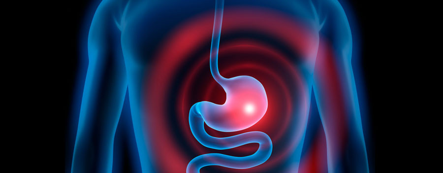
Endoscopy (stomachoscopy, colonoscopy, inspection of the biliary tract) means looking at the internal organs. By using most recent video endoscopes, we are able to have a closer look at the mucosae of the oesophagus, the stomach, the duodenum and the colon. As a result, we are able to locate abnormal mutations of the mucosae such as inflammations, ulcers, benign and malignant neoformations and bleedings.
In addition to that, we are able to take a biopsy via the endoscope. Following the biopsy, the tissue sample is sent to a pathologist in order to be examined under a microscope. The evaluation of the results usually takes 3 to 5 days.
I carry out outpatient and inpatient gastroscopies and coloscopies both for diagnosis and for therapy.
Adenoids throughout the gastrointestinal tract can be removed for therapeutic purposes, constrictions in the upper and lower gastrointestinal tract (e.g. after surgeries or chronic inflammatory bowel diseases) are extended, benign and malignant tumors are removed extensively, esophageal varices and fundus varices are atrophied and the PEG-system for an artificial nutrition can be placed.
When combining a sonographic unit with an endoscope, a device is created which allows the production of high-resolution images of the internal organs. By using this method, it is not only possible to isolate mucosal changes (for instance a swelling) and space-occupying processes (such as cysts and malignant tumors), but also to extract tissue samples for the pathologist.
Contact
Prof. Dr. med. R. J. Adamek
St. Josef-Krankenhaus Kupferdreh
Klinik für Innere Medizin
Heidbergweg 22-24
45257 Essen
Phone: +49 (0201) 455-1601
Fax: +49 (0201) 455-2959
E-Mail: r.adamek@contilia.de
Certificates